What is the normal anatomy and function of the hip joint?
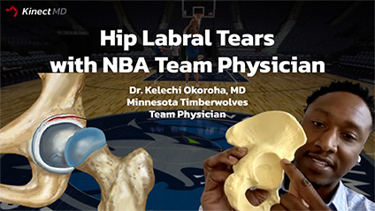
The hip is a ball and a socket joint that connects your torso or your trunk to your lower extremities. It is important to start the discussion with defining key anatomic features of the hip. This is the hemi pelvis of the hip which is half the hip joint. The hip or the pelvis bone is made up of three key bones you have the ilium which is the posterior portion you have the ischium which is the inferior portion and you have the pubis which is the anterior superior portion. In the center of these three bones is the acetabulum which is the socket of the hip and where the femoral head articulates. On the other side is your femur or your thigh bone and at the end of the femur is the femoral head. The femoral head is what articulates with that acetabulum. On the surface of the femoral head and the acetabulum is what we call the cartilage and the cartilage is the skin of the bones and it helps to provide a smooth gliding surface for those two bones and also assist in reducing compressive forces. As you watch the femoral head articulating with the acetabulum you'll notice that the acetabulum does not fully cover the femoral head, so in order to increase the coverage of the femoral head we have the Labrum. The labrum is a horseshoe structure that lines the acetabulum it functions to increase the volume of the hip. The labrum increases the volume of the hip by 33 percent. It also increases the contact area about 22 percent. Why is contact area important? i'll give you an example, if two people are carrying a 500 pound load that's going to be quite heavy for the two people to carry but if you have 10 people carrying the 500 pound load it makes it a lot easier for them to carry. Similarly, if you increase that contact surface area by 22 that's more surface area to distribute that load of your body weight. Also inside the hip is the synovial fluid and the synovial fluid is the natural fluid inside the joint and it provides nutrition to the articular cartilage or the skin that we talked about and allows that smooth gliding surface and also allows distribution of forces. If there is a labrum tear some of that fluid can leak out and that decreases the nutrition to your cartilage and that decreases the distribution of forces.
What is the pathophysiology of these hip label tears?
Labral tears can happen in multiple different situations. They can occur from traumatic injury , for example an athlete who has a hip dislocation or a subluxation however that occurs less commonly. More commonly there is a bony abnormality, either a patient has femoral acetabular impingement (FAI) where there is extra bone on either that femoral head/neck or that socket or there is hip dysplasia, where there is not enough coverage of that femoral head on the acetabulum.
Another function of the labrum is it acts as a suction seal and resists distraction of the femoral head from the socket. The cartilage works well in dispersing compressive loads however it doesn’t function well against shear forces. So if that hip is unstable and the femoral head is gliding in and out that's causing shear forces on the cartilage, that can lead to cartilage wear and arthritis.
How do patients with hip labral tears present?
Most of the time they complain of a chronic constant dull pain, however there can be periods where that's sharp and that's usually when they are active for instance extended walking, pivoting, prolonged sitting and impact activities. Some patients can describe some night pain but often symptoms have been present for a long time
Are there any demographic factors that increase hip labral tear incidence?
There is no difference in tear rates between males and females. However, what you'll notice is that as people get older the incidence of labral tears increases similar to what we see with meniscus tears. We have found in our studies that a large percentage of people have what we call asymptomatic label tears. If you obtain an mri of everyone over the age of 40 or 50 a high percentage of people will have a label tear. As you get older and the cartilage and the labrum degenerate you're going to have more labral tearing. The question is who do we need to treat?
What are some of the associated conditions and how do you rule out the other conditions?
One of the most common associated conditions is femoroacetabular impingement. Femoroacetabular impingement is when there is extra bone either on the femoral neck or extra bone on the acetabulum. This causes increased contact of the bones at earlier range of motion and that can lead to further bony abnormalities or labral tearing. This is a common factor especially in our high impact athletes. It is developed because when patients are young and their growth plate is open and there is repetitive flexion activity the bones can grind on each other and that can increase bone formation on the hip.
How do you diagnose Hip Labral tears?
Similar to the examination maneuvers in with knee meniscus tears you want to increase the stress on the labrum and then apply some kind of twisting factor to see if it illicits pain or mechanical symptoms. One of the main tests is the FADIR test. Hip flexion, adduction, and internal rotation. You flex the hip up and internally rotate and what you're trying to do is increase contact between the femoral neck and that labrum. If there is a tear the pain receptors in the labrum are going to transmit that pain. This test is most sensitive in diagnosing anterior superior tears. Another test is hyper extension or abduction and that's going to identify your posterior label tears. Another test is the fitzgerald test where you have your patient's hip flexed and then you extend while internally rotating the hip again trying to make some contact between those bones and elicit pain with the labral tears
Audience question: What are your recommendations for lack of mobility or stability in the hips besides the standard exercises and what is the role of the core in this in this situation?
Lack of mobility and lack of stability are two different ends of the spectrum for me. Lack of mobility could be due to body impingement like in FAI syndrome and that's a problem that may not correct with physiotherapy depending on the degree. Lack of stability could be from the labral tear or acetabular dysplasia and that has surgical implications. The core is important in terms of treatment and we will get into that later but also important is hip range of motion, hip strengthening, abductors, adductors. those are all pretty important with physical therapy.
What imaging do you use to diagnose labral tears?
The basic imaging is going to be an x-ray. X-rays will tell you about any kind of bony abnormalities and so on x-ray is you can identify pincer lesions (extra bone on the acetabulum) and cam lesions (extra bone on the femoral neck). Those are key signs that there is some impingement going on which could lead to labral tears. Also you want to evaluate the lateral acetabulum. The labrum is a soft tissue structure so you can't see it on the x-ray but sometimes when you have degeneration you can have calcification in that labral tissue and you can pick that up on the x-ray.
Then you have your advanced imaging. There is ultrasound imaging and MRI. Ultrasound is going to assess the soft tissues, so any kind of muscular abnormalities that are going on, any hip strains, any effusion in the hip. With the ultrasound you really can't get a good look at the cartilage or the labrum and that's what an MRI allows you to evaluate. An MRA is when you inject contrast or fluid into the hip to increase your sensitivity in identifying those labral tears or cartilage injuries. MRI is the gold standard when evaluating labral tears.
Can you elaborate more on the difference between an MRA and an MRI?
Injecting contrast in the joint increases the sensitivity of the test because when there is a labral tear the contrast will come out of the hip joint and there can be cystic changes. That can identify a labral tear.
When testing for anterior label tears how can you tell the difference between pain from FAI and a labral tear?
That's a really good question, you really can't tell the difference because often they overlap. When there is FAI you have a bony abnormality and often times there is also a label tear. With those tests you're rubbing that bone into that labrum and causing pain. So, to answer your question you really can't differentiate between the two and they often overlap.
What are the conservative management options for Hip labral tears and FAI (Hip Impingement)?
I am a big proponent of conservative management especially because like I stated there is a high percentage of people who have asymptomatic label tears. So just because we identify a labral tear on MRI doesn't mean a person needs surgery. I will often recommend a course of physical therapy first. In therapy there is a focus on strengthening of the core and hip while also trying to improve range of motion. We also try to strengthen the abductors and adductors. Additionally, we recommend rest or activity modification with low impact activities to allow the hip to rest and allow inflammation resolution. Pain can be managed with non-steroidal anti-inflammatories and other pain medications. If a patient has an intense level of pain we can perform a steroid injection. An intra-articular injection does two things it's diagnostic in that if a patient gets relief from an injection inside the hip that tells us that the pain is really coming from inside the hip. It is also therapeutic because it allows pain relief and allows augmentation of physical therapy.
What are your indications for surgical management of Hip Labral tears?
My indications for surgery are failure of non-operative treatment. So, failure of 6 - 12 weeks of physical therapy, anti-inflammatories and activity modification. If they fail and they're still having pain they then I indicate them for surgery. Snother indication is any kind of mechanical symptoms, for instance catching or locking in the hip. That is a sign that there might be displaced tissue or cartilage that needs to be addressed. Another indication is instability in the hip. The hip cartilage is good in resisting compressive loads when the hip is stable. When the hip is unstable there can be shearing loads, which are not good for the cartilage. So hip instability is an indication for labral repair surgery.
You stated that often times patients have concurrent labral tears and FAI (hip impingement). Do you treat these two conditions at the same time during surgery?
Surgery is really a la carte. So, we're going to identify everything that’s abnormal in the hip and we address it at the same time. Hip arthroscopy is minimally invasive and can be performed through three small incisions and a small camera. We look for any anatomic deformities, we shave down the extra bone on the acetabulum (socket), we shave down the extra bone on the femoral neck (CAM) and we also repair that labrum using suture anchors.
Somebody asked what is the role of genetic disorders in labral tears?
Genetic orders such as Marfans syndrome and Ehlers Danlos Syndrome can lead to Hip instability. When the hip is unstable, there is decreased coverage of the femoral head and decreased contact area. This increases the stress on different areas of the bone and that could be a risk factor for labral tears and hip instability.
Is there an increased risk of hip arthritis after labral repairs?
That's a really good question and a lot of studies have tried to speculate whether hip labral repair decreases the risk of arthritis in the future. the short answer is we don't know yet. Hip arthritis takes many years to develop and we just don't have the research yet to answer that. But I my belief is that it probably does decreases the long term arthritis risk. If we are able to re-establish that suction steel you stabilize the hip and then there is less stress on the cartilage and less degeneration leading to less arthritis.
What are the return to sports rates after Hip Labral repair?
The return of sports rates are actually pretty high. We've done multiple studies evaluating return to sports rates in different sports and found anywhere from 80 to 98% RTS when looking at football, basketball, hockey and soccer. They all have a high return to sport rate. The return to sports time is usually at least six months.
What percent of hip labral tears do you debride vs. repair?
That's a great question, when we first started doing hip arthroscopy surgeons were doing a lot of label debridements because we didn't have the instruments or implants to really do labor repairs. Following these early surgeries what we found was that we thought that patients did well but we were assessing them with the wrong kind of outcomes score. We were looking at outcomes using scores made for patients with hip arthroplasty whereas these patients were young and active. The current literature demonstrates that labral preservation is superior to labral debridement, so I try to repair every labral tear that I can. if the labrum is degenerative or deficient then the patient is looking at a labral augmentation or a labor reconstruction procedure.
Can you speak on the association between hockey goalies and Hip Impingement?
Hockey goalies have a high incidence of femoral acetabular impingement due to their position, goalies are usually in a flexed hip position and repetitively move from that position to even higher degrees of hip flexion with high force. By performing these actions over and over again goalies can develop femoral cam or pincer deformities. They have a similar return to sports rate as other sports/positions after surgery when we are able to correct those deformities on the femoral neck and the acetabulum. These players return to sports anywhere from six to nine months .
What are the common sports we see Hip labral tears and Hip Impingement in?
I think it is actually more common than we think, however athletes don't usually present with pain and if they do they are able to manage it. When we get a lot of these x-rays in our professional players a lot of them have femoral cam deformities and they're doing just fine. They are just at such a higher level that they're able to adapt and function with hip impingment.
Are there any interesting cases or any professional players cases that you could discuss?
I will state as a caveat that we don't have any player’s imaging/exam and we don't know exactly how a player was treated so all we can do is speculate how a similar injury would be treated. We can't give an exact treatment regimen for any player.
For everybody we are discussing Isaiah Thomas who had a labral tear back in 2016-2017.
We presume he had a labral tear and we don't know what other components he had, but I’m assuming he had femoroacetabular impingement. He obtained various opinions and he saw multiple different doctors. I am sure many surgeons believe a player with a similar injury should have had surgery right away but conservative management was ultimately chosen and he did an extensive course of conservative management. Our recent literature has demonstrated that patients treated with conservative management longer than six months have worse outcomes after hip arthroscopy. After suffering from a labral tear, the hip functions differently. There is shear forces on the cartilage if there is instability and increased contact stresses that can lead to arthritic changes. That might not be bad in in our everyday person who's working out let's say once or twice a week but in an nba player that’s competing no that hip every day 82 games a year that constant stress and increased load can lead to a more rapid progression of arthritis. He ended up getting surgery and he didn't do so well after it. In my opinion he may have already developed some arthritis. Now he had a hip resurfacing procedure, so I presume he had some end stage arthritis and the cartilage was worn out and the surgeon resurfaced his femoral head. The procedure is similar to replacing half the hip and now he's doing well.
what sort of conversations are you having with the team strength and conditioning coach when taking the athlete through the return to play protocols to get them back into sports safely with max performance?
There are different phases to post-operative rehabilitation and I have my rehab protocols online on the website if anyone who wants to take a look at them.
Initially we want to act conservatively and reduce inflammation while beginning gentle range of motion. Patients begin stationary bike and stretching by the therapist and then we progress at different checkpoints. I am in constant contact with all the strength and conditioning coaches afterwards and we assess the patient together make sure they're progressing along our checkpoints in our protocol.
How common is it for professional athletes to have hip resurfacing?
It is not a very common because we like to get to these injuries before an athlete has severe arthritis. There have been a couple of professional athletes I am aware of that had hip resurfacing and returned and I.T. is one of them but it's not very common to answer your question.
How has COVID-19 changed heart health monitoring in athletes?
That's a very good question. It has changed monitoring a lot. We're a lot more conscious of heart health and the need to get echocardiograms and because as you stated a lot of these athletes with COVID have had myocarditis that we were surprised to find out about.
Do you notice any changes in gait with these symptomatic labral tear patients?
It depends on the pathology. Some patients can have what we call micro instability and the hip can sublux when they are walking. That is a gait pattern that we look for. Also patients can have abductor weakness and can have a subtle lurch.
Where is your post-operative Hip arthroscopy protocol posted?
It is listed on KelechiOkorohaMD.com under patient resources and physical therapy protocols.
What is the most satisfying thing about your job and the reason you went into orthopedics and sports medicine? Could you talk about your journey and your inspiration?
Knowing the passion and the love I had for sports, I know what it would feel like not to be able to play sports. So I understand when I have these athletes with big injuries like labral tears or ACLs and they think their career is shattered and they have that look in their eyes of defeat. For me getting those patients back to doing the things they love, the sports that they love playing is the biggest satisfaction I can get.
Can Femoral CAM lesions grow back?
The abnormal bone growth usually does not grow back. However following surgery there can be heterotopic ossification. That is why we place patient on NSAIDS afterwards but that is not really the femoral cam growing back, it is bony formation in the soft tissues around the hip. Usually if an appropriate CAM resection is performed it doesn't grow back.
Are retired athletes more susceptible to injuries and what injuries in particular do these athletes have?
I don't know if retired athletes per se are more susceptible to injuries but a lot of retired athletes have degenerative changes for instance degenerative meniscus tears, degenerative labral tears and those things can increase the contact stresses in the joint and lead to other injuries/arthritis. But I don't believe it's just because they're retired.


